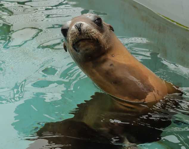
Loss of Neuron Connectors in the Brains of Sea Lions with Epilepsy
- Domoic acid
- Epilepsy
Abstract
One in 26 people develop epilepsy and in these temporal lobe epilepsy (TLE) is common. Many patients display a pattern of neuron loss called hippocampal sclerosis. Seizures usually start in the hippocampus but underlying mechanisms remain unclear. One possibility is insufficient inhibition of dentate granule cells. Normally parvalbumin‐immunoreactive (PV) interneurons strongly inhibit granule cells. Humans with TLE display loss of PV interneurons in the dentate gyrus but questions persist. To address this, we evaluated PV interneuron and bouton numbers in California sea lions (Zalophus californianus) that naturally develop TLE after exposure to domoic acid, a neurotoxin that enters the marine food chain during harmful algal blooms. Sclerotic hippocampi were identified by the loss of Nissl‐stained hilar neurons. Stereological methods were used to estimate the number of granule cells and PV interneurons per dentate gyrus. Sclerotic hippocampi contained fewer granule cells, fewer PV interneurons, and fewer PV synaptic boutons, and the ratio of granule cells to PV interneurons was higher than in controls. To test whether fewer boutons was attributable to loss versus reduced immunoreactivity, expression of synaptotagmin‐2 (syt2) was evaluated. Syt2 is also expressed in boutons of PV interneurons. Sclerotic hippocampi displayed proportional losses of syt2‐immunoreactive boutons, PV boutons, and granule cells. There was no significant difference in the average numbers of PV‐ or syt2‐positive boutons per granule cell between control and sclerotic hippocampi. These findings do not address functionality of surviving synapses but suggest reduced granule cell inhibition in TLE is not attributable to anatomical loss of PV boutons.
Cameron, S., Lopez A., Glabman, R., Abrams, E., Johnson, S., Field, C., Gulland, F.M., Buckmaster, P.S. 2019. Proportional loss of parvlbumin-immunoreacitve synaptic boutons and granule cells from the hippocampus of sea lions with temporal lobe epilepsy. J. Comp Neurology Mar. 12 doi:10.1002/cne.24680
Meet The Experts
{"image":"\/People\/Portrait\/cara-field.jpg","alt":"Cara Field","title":"Cara Field","text":"Director, Conservation Medicine","link_url":"https:\/\/www.marinemammalcenter.org\/person\/cara-field","link_text":"Read Bio"}

Related Publications
{"image":"\/Animals\/Patients\/California sea lions\/cropped-images\/csl-eeg-frances-gulland-photo-c-the-marine-mammal-center-33-0-806-630-1685576701.jpg","alt":"","title":"Pinniped Electroencephalography: Methodology and Findings in California sea lions","link_url":"https:\/\/www.marinemammalcenter.org\/publications\/pinniped-electroencephalography-methodology-and-findings-in-california-sea-lions","label":"Research Paper"}
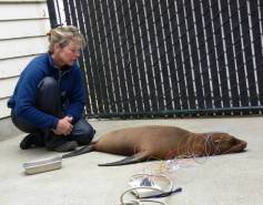
Pinniped Electroencephalography: Methodology and Findings in California sea lions
Read MoreRecent News
{"image":"\/Misc\/Graphics\/michelle-oates-detkin-graphic-1.png","alt":"Michelle Oates Detkin","title":"The Marine Mammal Center Welcomes New Board Member Michelle Oates Detkin","link_url":"https:\/\/www.marinemammalcenter.org\/news\/the-marine-mammal-center-welcomes-new-board-member-michelle-oates-detkin","label":"News Update","date":"2026-07-15 10:53:00"}
The Marine Mammal Center Welcomes New Board Member Michelle Oates Detkin
July 15, 2026
Read More{"image":"\/Animals\/Patients\/Hawaiian monk seals\/2022\/hmsahonuireleasephoto-1-by-megan-ely-c-noaanoaa-permit-18786.png","alt":"Hawaiian monk seal release","title":"Take Action: Proposed Federal Regulation Would Weaken Scientific Funding and Integrity","link_url":"https:\/\/www.marinemammalcenter.org\/news\/take-action-proposed-federal-regulation-would-weaken-scientific-funding-and-integrity","label":"News Update","date":"2026-06-30 09:24:00"}

Take Action: Proposed Federal Regulation Would Weaken Scientific Funding and Integrity
June 30, 2026
Read More{"image":"\/Animals\/Patients\/Steller sea lions\/2025\/sslwestley43photo-by-bill-hunnewell-c-the-marine-mammal-center-1.jpg","alt":"Steller sea lion, Westley.","title":"A Steller Story: Endangered Steller Sea Lion Westley Released in Alaska","link_url":"https:\/\/www.marinemammalcenter.org\/news\/endangered-steller-sea-lion-westley-released-in-alaska","label":"Patient Update","date":"2026-06-18 02:00:00"}

A Steller Story: Endangered Steller Sea Lion Westley Released in Alaska
June 18, 2026
Read More{"image":"\/Animals\/Wild\/Gray whale\/gray-whale-angle-by-josie-slaathaug-c-the-marine-mammal-center.jpg","alt":"A gray whale in San Francisco Bay","title":"The Wall Street Journal: Huge, Hungry Whales Are San Francisco\u2019s Latest Traffic Headache","link_url":"https:\/\/www.marinemammalcenter.org\/news\/wall-street-journal-huge-hungry-whales-are-san-franciscos-latest-traffic-headache","label":"In the News","date":"2026-05-19 15:32:00"}
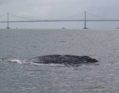
The Wall Street Journal: Huge, Hungry Whales Are San Francisco’s Latest Traffic Headache
May 19, 2026
Read More