Infection Dynamics of Influenza Virus A in California Sea Lions and Northern Elephant Seals
- Infectious disease
Abstract
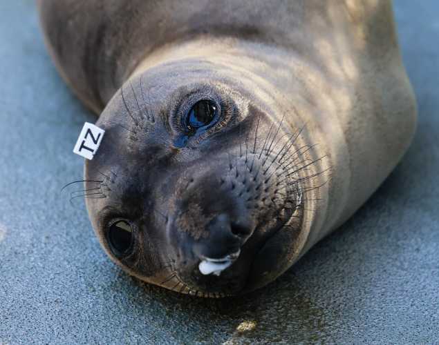
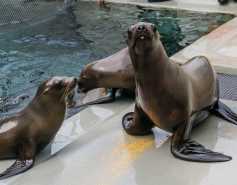
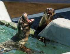
To understand susceptibility of wild California sea lions and Northern elephant seals to influenza A virus (IAV), we developed an ex vivo respiratory explant model and used it to compare infection kinetics for multiple IAV subtypes. We first established the approach using explants from colonized rhesus macaques, a model for human IAV. Trachea, bronchi, and lungs from 11 California sea lions, 2 Northern elephant seals, and 10 rhesus macaques were inoculated within 24 h postmortem with 6 strains representing 4 IAV subtypes. Explants from the 3 species showed similar IAV infection kinetics, with peak viral titers 48 to 72 h post-inoculation that increased by 2 to 4 log10 PFU/explant relative to the inoculum. Immunohistochemistry localized IAV infection to apical epithelial cells. These results demonstrate that respiratory tissue explants from wild marine mammals support IAV infection. In the absence of the ability to perform experimental infections of marine mammals, this ex vivo culture of respiratory tissues mirrors the in vivo environment and serves as a tool to study IAV susceptibility, host range, and tissue tropism. IMPORTANCE Although influenza A virus can infect marine mammals, a dearth of marine mammal cell lines and ethical and logistical challenges prohibiting experimental infections of living marine mammals mean that little is known about IAV infection kinetics in these species. We circumvented these limitations by adapting a respiratory tract explant model first to establish the approach with rhesus macaques and then for use with explants from wild marine mammals euthanized for nonrespiratory medical conditions. We observed that multiple strains representing 4 IAV subtypes infected trachea, bronchi, and lungs of macaques and marine mammals with variable peak titers and kinetics. This ex vivo model can define infection dynamics for IAV in marine mammals. Further, use of explants from animals euthanized for other reasons reduces use of animals in research.
Liu, H., Plancarte, M., Ball, E.E., Weiss, C.M., Gonzales-Viera, O., Holcomb, K., Ma, Z.M., Allen, A.M., Reader, J.R., Duignan, P.J. and Halaska, B., 2020. Respiratory tract explant infection dynamics of influenza A virus in California sea lions, northern elephant seals, and rhesus macaques. Journal of Virology, pp.JVI-00403.
Meet The Experts
{"image":"\/People\/Portrait\/cropped-images\/PD_Gray whale Pacifica_IMG_0552-0-0-1270-992-1775170630.jpeg","alt":"Padraig Duignan","title":"P\u00e1draig Duignan","text":"Senior Director, Research Pathology","link_url":"https:\/\/www.marinemammalcenter.org\/person\/padraig-duignan","link_text":"Read Bio"}
Related Publications
{"image":"\/Animals\/Wild\/Other species\/nz-sea-lion-shutterstock.jpg","alt":"New Zealand sea lion","title":"Causes of Death in Two Populations of New Zealand Sea Lions","link_url":"https:\/\/www.marinemammalcenter.org\/publications\/causes-of-death-in-two-populations-of-new-zealand-sea-lions","label":"Research Paper"}
{"image":"\/Animals\/Patients\/California sea lions\/csl-by-bill-hunnewell-c-the-marine-mammal-center-6.jpg","alt":"California sea lions","title":"Zoonotic Bacteria Persistence and Susceptibility","link_url":"https:\/\/www.marinemammalcenter.org\/publications\/zoonotic-bacteria-persistence-and-susceptibility","label":"Research Paper"}
{"image":"\/Animals\/Patients\/California sea lions\/cropped-images\/csl-photo-by-bill-hunnewell-c-the-marine-mammal-center-1-0-0-2358-1722-1600891644.jpg","alt":"California sea lions","title":"Emerging Viruses in Marine Mammals","link_url":"https:\/\/www.marinemammalcenter.org\/publications\/emerging-viruses-in-marine-mammals","label":"Research Paper"}
{"image":"\/Animals\/Patients\/California sea lions\/csl-photo-by-bill-hunnewell-c-the-marine-mammal-center-12.jpg","alt":"two California sea lions","title":"Multi-Phase Muscle Breakdown in California Sea Lions","link_url":"https:\/\/www.marinemammalcenter.org\/publications\/multi-phase-muscle-breakdown-in-california-sea-lions","label":"Research Paper"}
Recent News
{"image":"\/Misc\/Graphics\/michelle-oates-detkin-graphic-1.png","alt":"Michelle Oates Detkin","title":"The Marine Mammal Center Welcomes New Board Member Michelle Oates Detkin","link_url":"https:\/\/www.marinemammalcenter.org\/news\/the-marine-mammal-center-welcomes-new-board-member-michelle-oates-detkin","label":"News Update","date":"2026-07-15 10:53:00"}

The Marine Mammal Center Welcomes New Board Member Michelle Oates Detkin
July 15, 2026
Read More{"image":"\/Animals\/Patients\/Hawaiian monk seals\/2022\/hmsahonuireleasephoto-1-by-megan-ely-c-noaanoaa-permit-18786.png","alt":"Hawaiian monk seal release","title":"Take Action: Proposed Federal Regulation Would Weaken Scientific Funding and Integrity","link_url":"https:\/\/www.marinemammalcenter.org\/news\/take-action-proposed-federal-regulation-would-weaken-scientific-funding-and-integrity","label":"News Update","date":"2026-06-30 09:24:00"}

Take Action: Proposed Federal Regulation Would Weaken Scientific Funding and Integrity
June 30, 2026
Read More{"image":"\/Animals\/Patients\/Steller sea lions\/2025\/sslwestley43photo-by-bill-hunnewell-c-the-marine-mammal-center-1.jpg","alt":"Steller sea lion, Westley.","title":"A Steller Story: Endangered Steller Sea Lion Westley Released in Alaska","link_url":"https:\/\/www.marinemammalcenter.org\/news\/endangered-steller-sea-lion-westley-released-in-alaska","label":"Patient Update","date":"2026-06-18 02:00:00"}

A Steller Story: Endangered Steller Sea Lion Westley Released in Alaska
June 18, 2026
Read More{"image":"\/Animals\/Wild\/Gray whale\/gray-whale-angle-by-josie-slaathaug-c-the-marine-mammal-center.jpg","alt":"A gray whale in San Francisco Bay","title":"The Wall Street Journal: Huge, Hungry Whales Are San Francisco\u2019s Latest Traffic Headache","link_url":"https:\/\/www.marinemammalcenter.org\/news\/wall-street-journal-huge-hungry-whales-are-san-franciscos-latest-traffic-headache","label":"In the News","date":"2026-05-19 15:32:00"}
The Wall Street Journal: Huge, Hungry Whales Are San Francisco’s Latest Traffic Headache
May 19, 2026
Read More