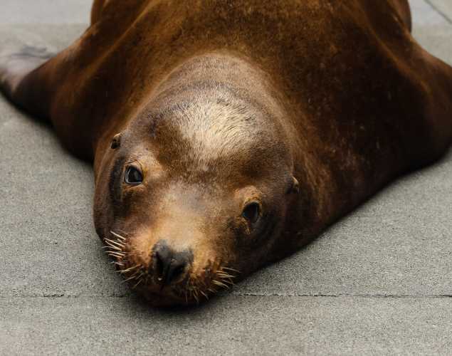
Postmortem Imaging Reveals Altered Hippocampal Connectivity in Sea Lions with Chronic Domoic Acid Toxicosis
- Domoic acid
- Pathology
- Diagnostics
Abstract
Hundreds of wild California sea lions (Zalophus californianus) exposed to the algal neurotoxin domoic acid are treated in veterinary rehabilitation centers each year. Common chronic effects of toxic exposure in these animals are seizures and hippocampal damage, and they have been proposed as a natural animal model for human epilepsy. Humans with medial temporal lobe epilepsy present with white matter pathology in a number of tracts including the fornix and increased structural connectivity between the hippocampus and thalamus. However, there are no prior data on structural connectivity in sea lion brains, with or without neuropathology. In the present study, we used a novel diffusion tensor imaging technique to obtain high resolution (1mm isotropic) white matter maps in brains obtained opportunistically postmortem from wild sea lions with and without chronic clinical signs of toxic exposure to domoic acid. All animals had received a full veterinary workup and diagnosis prior to euthanasia. We measured hippocampal atrophy morphometrically, and all brains were examined histopathologically. In animals diagnosed with chronic domoic acid toxicosis, the fornix showed signs of altered diffusion properties indicative of pathology; these brains also had increased structural connectivity between hippocampus and thalamus in comparison to brains from animals with no neurological signs. These findings establish further parallels between human medial temporal lobe epilepsy and a naturally occurring condition in wild sea lions and simultaneously advance general knowledge of the deleterious effects of an increasingly common natural toxin.
Cook, P.F., Berns, G.S., Colegrove, K., Johnson, S., Gulland, F. 2017. Post-mortem DTI reveals altered hippocampal connectivity in wild California sea lions. The Journal of Comparative Neurology Research in Systems Neuroscience. doi: 10.1002/cne.24317.
Related Publications
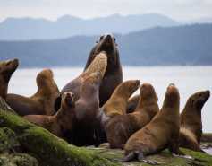
{"image":"\/Animals\/Patients\/California sea lions\/2020\/csl-oso-by-bill-hunnewell-c-the-marine-mammal-center-1.jpg","alt":"California sea lion","title":"Loss of Neuron Connectors in the Brains of Sea Lions with Epilepsy","link_url":"https:\/\/www.marinemammalcenter.org\/publications\/loss-of-neuron-connectors-in-the-brains-of-sea-lions-with-epilepsy","label":"Research Paper"}
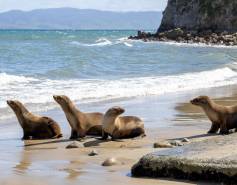
{"image":"\/Animals\/Wild\/California sea lion\/cropped-images\/sea-lion-pup-shutterstock-451-142-3018-2358-1603915228.jpg","alt":"California sea lion pup","title":"Domoic Acid in Fetal Fluids of California Sea Lions","link_url":"https:\/\/www.marinemammalcenter.org\/publications\/domoic-acid-in-fetal-fluids-of-california-sea-lions","label":"Research Paper"}
{"image":"\/Animals\/Patients\/California sea lions\/cropped-images\/csl-by-bill-hunnewell-c-the-marine-mammal-center-2-0-1031-1794-1401-1603916128.jpg","alt":"California sea lion","title":"Evaluating Sense of Smell in California Sea Lions and Relevance to Domoic Acid","link_url":"https:\/\/www.marinemammalcenter.org\/publications\/evaluating-sense-of-smell-in-california-sea-lions-and-relevance-to-domoic-acid","label":"Research Paper"}
Evaluating Sense of Smell in California Sea Lions and Relevance to Domoic Acid
Read More{"image":"\/Animals\/Wild\/Steller sea lion\/cropped-images\/steller-sea-lion-harem-shutterstock-648-0-4424-3456-1603916879.jpg","alt":"Group of Steller sea lions","title":"Algal Toxins in Alaskan Marine Mammals Foraging in a Changing Environment","link_url":"https:\/\/www.marinemammalcenter.org\/publications\/algal-toxins-in-alaskan-marine-mammals-foraging-in-a-changing-environment","label":"Research Paper"}
Related News
{"image":"\/Animals\/Wild\/California sea lion\/cropped-images\/csl-release-4-5-24photo-by-chris-deimler-c-the-marine-mammal-center-138-0-1270-992-1745348188.jpg","alt":"Four young California sea lions walk on the beach toward the ocean.","title":"Achievements in Ocean Health","link_url":"https:\/\/www.marinemammalcenter.org\/news\/achievements-in-ocean-health","label":"News Update","date":"2025-04-23 10:20:00"}
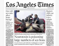
{"image":"\/Animals\/Patients\/California sea lions\/cropped-images\/LA Times Front Page 8.13.24-340-15-1270-992-1723588213.png","alt":"LA Times Front Page Domoic Acid Sea Lions","title":"LA Times: Neurotoxin is Poisoning Large Numbers of Sea Lions","link_url":"https:\/\/www.marinemammalcenter.org\/news\/la-times-neurotoxin-is-poisoning-large-numbers-of-sea-lions","label":"In the News","date":"2024-08-13 02:00:00"}
{"image":"\/Animals\/Patients\/California sea lions\/2024\/cropped-images\/musty - csl - bill hunnewell-180-0-1270-992-1723649438.jpg","alt":"California sea lion being treated for domoic acid poisoning","title":"NBC Nightly News: Researchers Investigate Dozens of Sick Sea Lions Along California Coast","link_url":"https:\/\/www.marinemammalcenter.org\/news\/nbc-nightly-news-researchers-investigate-dozens-of-sick-sea-lions-along-california-coast","label":"In the News","date":"2024-08-13 02:00:00"}
NBC Nightly News: Researchers Investigate Dozens of Sick Sea Lions Along California Coast
August 13, 2024
Read More{"image":"\/Animals\/Patients\/Releases\/cropped-images\/csl-quly-left-and-togozees-release-by-bill-hunnewell-c-the-marine-mammal-center-90-0-3996-3122-1693421090.jpg","alt":"two California sea lions are released in front of a crowd of people","title":"NPR Morning Edition: Hundreds of Seals and Sea Lions Are Treated Each Year at The Marine Mammal Center","link_url":"https:\/\/www.marinemammalcenter.org\/news\/npr-morning-edition-hundreds-of-seals-and-sea-lions-are-treated-each-year-at-the-marine-mammal-center","label":"In the News","date":"2023-08-18 02:00:00"}
NPR Morning Edition: Hundreds of Seals and Sea Lions Are Treated Each Year at The Marine Mammal Center
August 18, 2023
Read More